LECTURE
1 CPD
OCT in diabetic eye disease - Topcon
This session is now fully booked
About the session
CPD ref: C-107460
Description
OCT has enabled us to visualise the posterior and anterior segments for many different eye conditions and eye disease over a period of time. OCT has a massive benefit for clinicians with the visualisation of Diabetic Retinopathy, using this non-invasive technology. The progression of OCT-A gives clinicians further detailed information of the eye’s vascular network, further improving the ability to monitor diabetic retinopathy and maculopathy. The aim of this lecture is to show and talk through examples of different Diabetic pathology using OCT & OCTA to enable clinicians to understand the clinical signs of DR visible on OCT, the benefits of using OCT in practice to aid and help with clinical monitoring, decisions and referrals.
Target audience
- Optometrist
- Dispensing Optician.
Learning outcomes
Clinical practice
- Keep your knowledge and skills up to date - using the latest technology in OCT and OCT-A to assess and manage diabetic retinopathy
- Recognise, and work within, your limits of competence
- Conduct appropriate assessments, examinations, treatments and referrals - particular in respect to managing diabetic retinopathy.
Communication
- Listen to patients and ensure they are at the heart of decisions made about their care
- Communicate effectively with patients.
Speaker

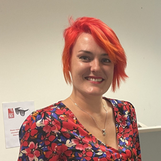
Danielle Lee

Dani has recently taken on the management role of the Clinical Affairs team after joining Topcon a few years ago, and has become an expert in our products, especially the Topcon MYAH, for myopia management and dry eye.
Dani graduated with a degree in Medical Biochemistry from the University of Birmingham and before joining Topcon spent seven years working in a number of roles across the Bristol Eye Hospital, primarily between the Imaging and Research departments.
Experienced in all aspects of ophthalmic imaging – including OCT, colour, infrared and autofluorescence photography, fluorescein and ICG angiography, OCT Angiography, Corneal topography, specular microscopy, handheld ERG & microperimetry as well as assisting in aseptic intravitreal procedures, checking visual acuity and intraocular pressure.
Dani also worked as a Clinical Trial Coordinator which involved the set-up, running, and maintenance of a variety of different ophthalmic trials of different phases, as well as certifying as an Imager for a variety of studies, allowing her to capture/process images required for study outcome assessments, and put in to place training plans and protocols.
