Pigmented fundus lesions

/COO/media/Media/CMGs/Pigmented-Fundus-Lesions-1-Small-410x250.jpg
Typical naevus nasal in the right eye, with numerous drusen indicating longevity of the lesion.
/COO/media/Media/CMGs/Pigmented-Fundus-Lesions-2-Large-445x645.jpg
Atypical choroidal naevus in the left eye. The tumour diameter exceeds 4DD but does not have any other features that are suspicious of malignancy. The drusen on its surface indicate that the lesion is longstanding.
/COO/media/Media/CMGs/Pigmented-Fundus-Lesions-3-Small-274x250.jpg
Small choroidal melanoma in the left eye, with a diameter exceeding 4 DD and large clumps of orange pigment.
/COO/media/Media/CMGs/Pigmented-Fundus-Lesions-4-Small-223x250.jpg
Congenital Hypertrophy of the Retinal Pigment Epithelium located superotemporally in the right eye. Note the deep pigmentation, discrete margins, halo, and lacuna.

Aetiology

Pigmented lesions of the choroid are a relatively common finding in the course of a routine examination of the fundus. The overwhelming majority are choroidal naevi, which are benign with a low risk of malignant transformation. Congenital Hypertrophy of the Retinal Pigment Epithelium (CHRPE) has distinctive ophthalmoscopic features. Choroidal melanoma is a rare and life-threatening melanocytic cancer that can sometimes be difficult to distinguish from naevus.


Choroidal naevus

  • area of increased choroidal pigmentation

- approx. 20% within the macular region, 70% between macula and equator and 10% between equator and ora serrata

  • low malignant potential (annual rate of malignant transformation of a choroidal naevus estimated to be 1 in 8,845 in a white US population)
  • prevalence reported as 2-7%. Population studies in some ethnicities report a lower prevalence: Hispanics (2.7%), African Americans (0.6%)

Congenital Hypertrophy of the Retinal Pigment Epithelium (CHRPE)

  • benign congenital anomaly of the retinal pigment epithelium (RPE)
  • area of increased RPE hypertrophy and hyperpigmentation
  • single lesion or multiple grouped CHRPE (‘bear tracks)’
  • malignant transformation is extremely rare, although slight enlargement is observed in 46% of those followed up photographically for >3 years

Uveal melanoma

  • 90% are choroidal, 6% arise from the ciliary body and 4% from the iris
  • 1.3-8.6 cases per million per year in European-derived populations
  • rare in Asians and very rare in Africans
  • life-threatening ocular malignancy (risk of metastatic disease)
    • detection and treatment at earliest stage improves prognosis
  • risk of metastases related to:
    • size and location
    • cell type and histopathological features
    • genetic changes (chromosome 3 loss or BAP1 mutation)
  • outcome poor once metastasis occurs; 1 year survival in 10-15%
  • mean age at presentation 60 years. Can occur at any age but rare in
  • childhood

Predisposing factors

Choroidal naevus

  • prevalence increases with age
  • ethnicity: most common in white people; rare in black people

Choroidal melanoma

  • melanoma arises de novo or from pre-existing choroidal naevus
  • risk factors for choroidal melanoma include: light coloured irides, fair skin, inability to tan, congenital ocular melanocytosis (‘naevus of Ota’) (1:400 lifetime risk of uveal melanoma), uveal melanocytoma, the BAP1 tumour predisposition syndrome, and, rarely, neurofibromatosis
  • the role of sunlight is uncertain, as most UV light is filtered by the lens

Symptoms of pigmented fundus lesions

Choroidal naevi

  • rarely symptomatic. Blurred or distorted vision can occur with sub-foveolar naevi or when associated with sub-retinal fluid or choroidal neovascular membrane

CHRPE

  • asymptomatic

Choroidal melanoma

  • symptoms include: photopsia, blurred or distorted vision, floaters, or field loss. In 30% of patients, choroidal melanoma is detected (usually by optometrists) before symptoms develop

Signs of pigmented fundus lesions

Choroidal naevus

  • typical naevi are small, flat and grey (though some are amelanotic), with a featureless surface and no sub-retinal fluid
  • atypical naevi are larger and dome-shaped, with or without drusen and/or traces of sub-retinal fluid; orange pigment absent
  • drusen indicate chronicity so that their absence over a domed lesion is suspicious

CHRPE

  • solitary, flat, well-demarcated deeply-pigmented lesion (but may contain discrete non-pigmented areas called lacunae)
  • shape elliptical or irregular
  • may show a narrow non-pigmented line at margins of lesion
  • the CHRPE lesion may (extremely rarely) develop low-grade adenocarcinoma on its surface

Choroidal melanoma

  • small choroidal melanomas are distinguished from naevi by:
    • larger size (i.e., thickness >2 mm and/or largest basal diameter (LBD) >6mm [>4 disc diameters])
    • there is a size overlap between choroidal naevi and choroidal melanomas with the following relative frequencies:
      • LBD 5-6mm: 70 naevi for 1 melanoma
      • LBD >6-7mm: 10 naevi for 1 melanoma
      • LBD >7-8mm: 3 naevi for 1 melanoma
    • clumps of orange pigment (which are hyper-autofluorescent)
    • subretinal fluid, most easily seen on OCT
    • documented growth (which requires sequential colour photography)
  • larger choroidal melanomas are dome-shaped, with more extensive serous retinal detachment. If they break through Bruch’s membrane and RPE, they can develop a mushroom (‘collar stud’) shape, which is almost pathognomic for melanoma
  • about 5% of choroidal melanomas are diffuse (i.e. with a wide base but minimal thickening). These tumours are aggressive and often extend extra-ocularly by the time of diagnosis
  • vitreous haemorrhage is rare, occurring only if the tumour has perforated the retina
  • the MOLES scoring system (see below) scores five features of melanocytic choroidal tumours that are suggestive of malignancy: mushroom shape, orange pigment, large size, enlarging tumour and sub-retinal fluid. Each feature is scored 0, 1, or 2 depending on whether they are absent, borderline/uncertain, or present. Tumours are diagnosed according to the sum of these five scores as: 
    - a common naevus (MOLES score = 0) 
    - low-risk naevus (score = 1) 
    - high- risk naevus (score = 2) 
    - probable melanoma (score 3 or more).
  • MOLES has been validated in an ocular oncology setting and shows a high sensitivity for indicating malignancy.

Differential diagnosis

Peripheral exudative haemorrhagic chorioretinopathy (‘eccentric disciform’)

Circumscribed choroidal haemangioma

Haemorrhagic detachment of RPE or retina

Age-related macular degeneration

Choroidal metastasis from cancer of lung, breast or other organs

Management by optometrist

Practitioners should recognise their limitations and where necessary seek further advice or refer the patient elsewhere 

Non pharmacological 

Dilated slit-lamp indirect biomicroscopy 

Baseline colour photography is essential, and sequential colour photography
is ideal, for demonstrating or excluding growth of the lesion

  • if imaging not possible, make careful drawing with measurement,
    using landmarks such as retinal blood vessels and optic disc
  • it is recommended that the patient be given a photograph, or access to a digital image, in case the next optometric assessment takes place elsewhere

OCT and autofluorescence imaging help to differentiate large naevi from small choroidal melanomas by demonstrating subretinal fluid and lipofuscin
(orange pigment) respectively

(GRADE*: Level of evidence=low, Strength of recommendation=strong)

Pharmacological 

None

Management category

A3 (modified): Probable melanoma. Urgent (within two weeks, in accordance with the Suspected CANcer pathway, SCAN) referral to ophthalmologist (RCOphth guidance):

  • patients with a suspicious melanocytic choroidal tumour having
    (A) any one of the following:
    • thickness greater than 2.0mm (or LBD greater than 7mm)
    • collar stud configuration
    • documented growth
  • or (B) any two of the following:
    • thickness greater than 1.5mm (or LBD greater than 6mm)
    • orange pigment
    • serous retinal detachment
    • symptoms
  • Alternatively, use the MOLES scoring system
    - MOLES score ≥3 is defined as “probable melanoma” 
    - local pathways defining referral criteria based on MOLES have been developed and should be followed where available

The MOLES scoring system for categorising melanocytic choroidal tumours according to likelihood of malignancy

IndicatorFindingScore
Mushroom shape

0 = Absent

1 = Incipient (erosion through RPE) / uncertain)

2 = Present (i.e. definitive mushroom shape with overhang)

 
Orange pigment

0 = Absent

1 = Dusting / unsure

2 = Confluent (i.e. easily visible clumps of orange pigment)

 
Large size

0 = Flat (<1mm thick) and less than 3 disc diameters (DD) wide

1 = Subtle dome shape (1-2mm thick) AND/OR 3- 4DD wide

2 = Significant thickening (>2mm) AND/OR more than 4DD wide

 
Enlargement

0 = None (or no baseline photography)

1 = Suspected change on comparing photographs

2 = Definite growth confirmed by sequential imaging

 
Subretinal fluid

0 = Nil

1 = Trace (limited retinal detachment seen only with OCT)

2 = Definite subretinal fluid visible with ophthalmoscopy

 
 Moles total score = 


B1 (modified): Routine referral to ophthalmologist followed by regular surveillance, which may be shared with ophthalmologist in accordance with local protocols. Where available, refer to local pathway:

  • low or high risk naevus
    • greater than 6mm LBD and dome-shaped, with or without drusen and/or traces of subretinal fluid; orange pigment absent (atypical naevus)

B3 (modified): inform the patient but no need for regular surveillance (review at subsequent routine eye examinations):

  • typical (common) naevus
    • none of the ‘high-risk’ features mentioned above
    • MOLES total score=0
    • applies even if lesion not previously documented
  • CHRPE
    • adenomas and adenocarcinomas associated with CHRPE are exceedingly rare
    • there is a rare but important association with Familial Adenomatous Polyposis (FAP), an inherited cancer predisposition syndrome, especially when the signs occur in multiple quadrants or are bilateral

Possible management by ophthalmologist

Choroidal melanoma

  • managed by a supra-regional multidisciplinary team
  • if diagnosis uncertain, options for indeterminate lesions are:
    • close observation in an ocular oncology clinic
    • fine needle aspiration biopsy for cytology, though sampling error may confound the diagnosis
    • genetic analysis (e.g. multiplex ligation-dependent probe amplification, or more recently next generation sequencing) determines whether the melanoma has metastatic potential
  • treatment dependent on tumour size and location and includes:
    • brachytherapy with a radioactive plaque
    • proton beam radiotherapy
    • laser therapy often as an adjunct to radiotherapy
    • surgical resection, trans-sclerally or trans-retinally
    • enucleation
  • postoperative monitoring for ocular complications such as local tumour recurrence, macular oedema, retinal detachment, neovascularisation and glaucoma as well as systemic surveillance for metastases

Evidence base

*GRADE: Grading of Recommendations Assessment, Development and Evaluation (www.gradeworkinggroup.org)

Sources of evidence

Al Harby L, Sagoo MS, O'Day R, Hay G, Arora AK, Keane PA, Cohen VM, Damato B. Distinguishing choroidal nevi from melanomas using the MOLES algorithm: Evaluation in an ocular nevus clinic. Ocul Oncol Pathol. 2021;7(4):294-302.

Augsburger JJ, Corréa ZM, Trichopoulos N, Shaikh A. Size overlap between benign melanocytic choroidal nevi and choroidal malignant melanomas. Invest Ophthalmol Vis Sci. 2008;49:2823-8

Damato B, Heimann H. Personalized treatment of uveal melanoma. Eye (Lond). 2013;27(2):172-9

Damato EM, Damato BE. Detection and time to treatment of uveal melanoma in the United Kingdom: an evaluation of 2,384 patients. Ophthalmology. 2012;119(8):1582-9

Damato B, Eleuteri A, Taktak AF, Coupland SE. Estimating prognosis for survival after treatment of choroidal melanoma. Prog Retin Eye Res. 2011;30(5):285-95

Damato BE. Can the MOLES acronym and scoring system improve the management of patients with melanocytic choroidal tumours? Eye (Lond). 2022:1-7. doi: 10.1038/s41433-022-02143-x. Online ahead of print.

Flanagan JP, O'Day RF, Roelofs KA, McGuinness MB, van Wijngaarden P, Damato BE. The MOLES system to guide the management of melanocytic choroidal tumours: can optometrists apply it? Clin Exp Optom. 2022:1-5. doi: 10.1080/08164622.2022.2029685. Online ahead of print.

Kaliki S, Shields CL. Uveal melanoma: relatively rare but deadly cancer. Eye (Lond). 2017;31(2):241-57

Nicholson BD, Oke J, Smith CF, Phillips J-A, Lee J, Abel L, Kelly S, Gould I, Mackay T, Kaveney Z, Anthony S, Hayles S, Lasserson D, Gleeson F. The Suspected CANcer (SCAN) pathway: protocol for evaluating a new standard of care for patients with non-specific symptoms of cancer. BMJ Open 2018;8:e018168

Mackay T, Kaveney Z, Anthony S, Hayles S, Lasserson D, Gleeson F. The Suspected CANcer (SCAN) pathway: protocol for evaluating a new standard of care for patients with non-specific symptoms of cancer. BMJ Open 2018;8:e018168

Roelofs KA, O'Day R, Harby LA, Arora AK, Cohen VML, Sagoo MS, Damato B. The MOLES System for Planning Management of Melanocytic Choroidal Tumors: Is It Safe? Cancers (Basel). 2020;12(5):1311

Royal College of Ophthalmologists. Referral pathways for adult ocular tumours March 2019

Shields CL, Manalac J, Das C, Ferguson K, Shields JA. Choroidal melanoma: clinical features, classification, and top 10 pseudomelanomas. Curr Opin Ophthalmol. 2014;25(3):177-85

Shields CL, Furuta M, Berman EL, Zahler JD, Hoberman DM, Dinh DH, Mashayekhi A, Shields JA. Choroidal nevus transformation into melanoma: analysis of 2514 consecutive cases. Arch Ophthalmol. 2009;127(8):981-7

Shields CL, Furuta M, Mashayekhi A, Berman EL, Zahler JD, Hoberman DM, Dinh DH, Shields JA. Clinical spectrum of choroidal nevi based on age at presentation in 3422 consecutive eyes. Ophthalmology. 2008;115(3):546-52

Shields CL, Mashayekhi A, Ho T, Cater J, Shields JA. Solitary congenital hypertrophy of the retinal pigment epithelium: clinical features and frequency of enlargement in 330 patients. Ophthalmology. 2003;110(10):1968-76

Singh AD, Kalyani P, Topham A. Estimating the risk of malignant transformation of a choroidal nevus. Ophthalmology. 2005;112(10):1784-9

Summary

What are Pigmented Fundus Lesions?

During an eye examination, optometrists look at the inside of the eye to make sure it is healthy. Sometimes they spot changes that might need monitoring or more tests.

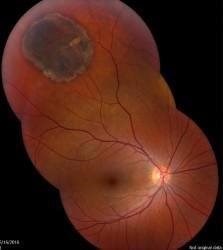
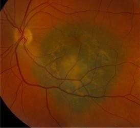
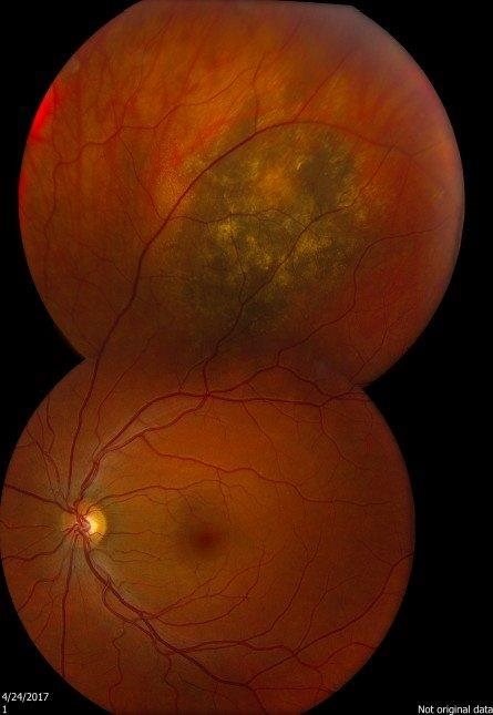
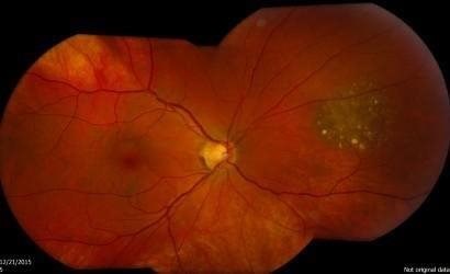
One change that is quite often seen is called a choroidal naevus. This is like a mole on the skin and is found in a part of the eyeball called the choroid. The choroid is the layer of nourishing and supportive tissue at the back of the eye between the retina (the part of the eye that receives light and sends images to the brain) and the outer coat of the eye. A choroidal naevus cannot be seen from the outside of the eye and usually causes no symptoms. Without professional eye examinations, people may never know they have one. If a choroidal naevus is found, the optometrist will check it at each regular eye examination.

If your choroidal naevus does not look normal, it is called an atypical choroidal naevus. This will be monitored by your ophthalmologist or optometrist through regular eye examinations as there is a small chance it may change into a cancer. They will take photographs of the inside of your eye so that they can see if your choroidal naevus changes or grows over time.

Some people are born with a harmless blemish inside the eye called Congenital Hypertrophy of the Retinal Pigment Epithelium (CHRPE). This can be confused with a choroidal naevus, but will not need to be regularly monitored.

Sometimes the optometrist will see a change inside the eye that might be a cancer called choroidal melanoma. Choroidal melanoma is very rare.

How are Pigmented Fundus Lesions managed?

If an optometrist thinks someone might have developed choroidal melanoma, they will refer that person to an ophthalmologist (a specialist eye doctor) within two weeks. The ophthalmologist will examine the eye and carry out some tests to see if there is a cancer.

A person diagnosed with choroidal melanoma will be offered treatment to reduce the risk of it spreading to other parts of the body and to save as much of the vision as possible. The treatment will depend on the type of cancer that is found and how advanced it is. More information about choroidal melanoma and different treatments can be found on Macmillan Cancer Support’s website.

Pigmented fundus lesions
Version 5
Date of search 14.03.23
Date of revision 27.04.23
Date of publication 12.06.23
Date for review 13.03.25
© College of Optometrists