Pterygium

A pterygium is a raised, triangular thickening of the bulbar conjunctiva that may extend onto part of the cornea. Prevalence is greater in males, older people and those with a history of long-term exposure to UV radiation. It can cause dryness, redness, foreign body sensation, inflammation, astigmatism and can threaten the visual axis. This Clinical Management Guideline outlines the aetiology, signs and symptoms and evidence-based recommendations for diagnosing and treating pterygium. It provides recommendations for non-pharmacological management and pharmacological treatment approaches, and for when referral may be appropriate.

Login to read further information on the diagnosis and management of pterygium.

/COO/media/Media/CMGs/Pterygium_001_BLURRED.jpg

What is pterygium?

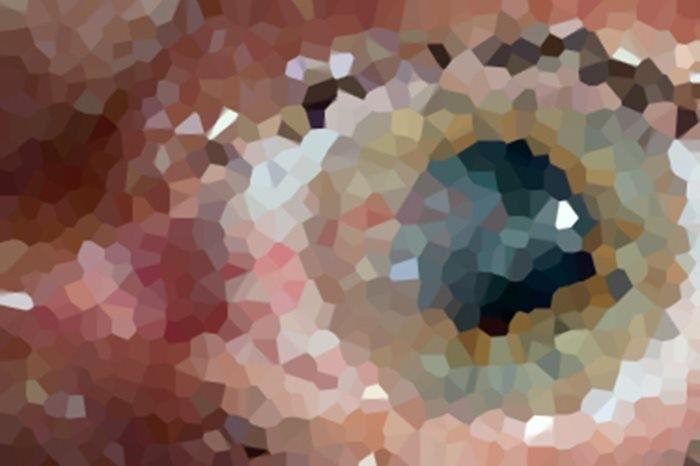
A pterygium is a triangular thickening of the conjunctiva (the layer of transparent skin on the white of the eye) which extends on to the outer edge of the cornea (the transparent window at the front of the eye).  If the cornea is imagined as a clock face, a pterygium normally occurs at the three and nine o’clock positions, more usually on the side nearest to the nose.  A pterygium may grow over the corneal surface. Because of tissue shrinkage, it can put tension on the cornea, causing astigmatism (loss of spherical curvature) and reducing the sharpness of vision.  Patients may complain of irritation of the affected eye and they may be concerned about the cosmetic appearance.

Pterygium is caused by long-standing exposure of the eyes to strong sunlight (particularly ultra-violet (UV) light).  It is also more likely to occur in people who spend a lot of time outdoors in dry, dusty conditions. Because sunlight is much stronger in countries closer to the equator, pterygium is more common in those regions.

How is pterygium managed?

Having carefully examined the affected eye(s) the optometrist will measure the size of the pterygium for future comparison. Artificial tears and lubricating ointment may be enough to control the irritation of the eyes.  If the pterygium is inflamed, a short course of steroid eye drops may be prescribed.  If the pterygium continues to grow towards the centre of the cornea, threatening the vision of the eye, or if inflammation cannot be controlled, the patient will be referred to an ophthalmologist (specialist eye doctor).  If the pterygium needs to be removed there are several different surgical techniques available.

Sign in to continue

Forgotten password?
Register

Want to read the rest of this page?

If you are a member, login to view this page. If you are not a member, simply register to gain free access to the rest of this content.

Last updated

Pterygium - 5 June 2026
The aetiology section has been amended to provide updated epidemiological data on prevalence and geographic distribution for pterygium. The symptoms section has been amended to include the mechanisms where pterygium may cause blurred or reduced vision and the impact of pterygium on contact lens wearers. The signs section now includes descriptions for classifying the different subtypes of pterygium. The possible management section has been updated to include more commonly performed surgical procedures for pterygium and treatment options for recurrent cases.

Version 11
Date of search 15.03.26
Date of revision 27.02.26
Date of publication 05.06.26
Date for review 14.03.28
© College of Optometrists